
Journal of Advanced Biological Sciences
ISSN Print: N/A
ISSN Online: 3134-8823
About: Journal of Advanced Biological Sciences (JABS) is a peer-reviewed, open-access journal that aims to publish cutting-edge research and advancements in all areas of biological sciences. The journal serves as a platform for researchers, academicians, and professionals to contribute their scientific knowledge and foster innovation in biology-related fields.
Publisher Responsibility
Lumina Quest Publishing is responsible for providing the journal with the necessary publishing infrastructure, editorial support, production services, digital preservation, and dissemination of published content. The publisher supports transparent and ethical publishing practices and ensures that the journal’s publishing operations are conducted in accordance with applicable scholarly publishing standards.
The publisher is also responsible for maintaining the journal’s website, publication systems, archiving arrangements, and administrative support while respecting the independence of editorial decision-making.
Editorial Responsibility
The Editor-in-Chief and Editorial Board are responsible for maintaining the academic and scientific quality of the journal. The editorial team oversees the evaluation of submitted manuscripts, selection and coordination of peer reviewers, editorial assessment and decisions, and adherence to the journal’s scope and ethical standards.
Editorial decisions are made independently on the basis of academic quality, originality, methodological soundness, relevance, and ethical compliance, without inappropriate influence from the publisher, authors, reviewers, or commercial interests.
Editorial Independence
The editorial team maintains editorial independence in all decisions concerning manuscript evaluation, peer review, acceptance, revision, and rejection. The publisher does not interfere with the academic judgment of editors or reviewers.
Journal of Advanced Biological Sciences | Year 2025 | Volume 2 | Issue 2 | Pages 30-39
Clinical Evaluation of CA15-3, Hematological Indices, Liver and Kidney Function Tests and Electrolyte Disturbances in Breast Cancer Patients
Lubna Faisal Hussein 1*1Department of Biology, College of Science, University of Baghdad, Baghdad, Iraq
View PDF Download XML Download DOI XML DOI: 10.66590/jabs2025020207
Abstract
Objective: Breast Cancer (BC) is the most common cancer worldwide and is the second leading cause of cancer deaths among women, its incidence continues to escalate. The development of BC is a multi-step process involving multiple cell types, early diagnosis of BC is one of the best approaches to prevent this disease. Methods: This case-control study included sixty women with breast cancer as well as 20 healthy women. The cases were selected using simple random sampling from Al-Amal National Hospital for Cancer Management. The present study performed to assess the levels of some parameters in BC women versus healthy women, which including CA15-3 marker, hematological parameters (including WBSs, RBCs, Hb, Platelets and ESR), liver function tests that included Aspartate Aminotransferase (AST), Alanine Aminotransferase (ALT) and Alkaline Phosphatase (ALP) kidney function tests (urea and creatinine) and some electrolytes (Cl-, K+, Na+ mmol/L). Results: The mean and Standard Deviation (SD) of the age of breast cancer patients are 51.81±11.323, while for healthy individuals, they are 30.25±4.756. The results of the statistical analysis showed significant differences (p = 0.004). The classification of patients by age into three groups showed that 31% were in the 25-45 age group, 35% in the 46-56 age group and 33.3% in the 57-77 age group. Regarding the assessment of studied parameters, the present study showed a significant (p = 0.032) increase in the levels of CA15-3 in women with breast cancer versus healthy women. As for hematological parameters, the statistical analysis showed significant differences only in RBCs, Hb and ESR: RBCs and Hb were significantly decreased (p = 0.038, p = 0.000, respectively) in breast cancer patients compared with controls, while ESR was significantly increased (p = 0.000). In terms of liver function tests, only ALP showed a significant (p = 0.000) increase in women with breast cancer patients as compared with control women; while AST and ALT showed non-significant differences. Regarding the assessment of kidney function tests and electrolytes, the statistical analysis showed that only urea increased significantly (p = 0.054) in patients, while creatinine and all studied electrolytes showed non-significant differences between patients and the control group. CA15-3 shows a significant inverse correlation with WBCs in breast cancer patients and with K+ only. No other significant correlations were found with the other studied parameters in either patients or healthy individuals. Conclusion: The current findings highlight the multifactorial nature of BC pathophysiology, where hematological, biochemical and metabolic shifts come together to shape cancer progression and prognosis.
INTRODUCTION
Breast Cancer (BC) is the most frequently diagnosed cancer in women and one of the leading causes of cancer-related death in females. The recent global estimates reveal that breast cancer accounted for nearly 670,000 deaths in 2024, with incidence expected to increase to 3.2 million new cases every year by 2050 if the current trend persists [1,2,3]. While it was once predominantly seen as a disease of developed countries, more than half of BC diagnoses and two-thirds of BC deaths in 2020 occurred in less developed regions of the world [4,5].
The incidence of BC is robustly linked with human development. Human development index, which incorporate measures of life expectation, education, and wealth, regards as a more inclusive and efficient comparison among countries than income alone [6]. In general, worldwide burden of cancer occurrence and mortality is growing rapidly, which is driving by population aging and growth, along with changes in prevalence and distribution of the principal cancer risk determinant, which are correlated to socioeconomic development [7,8].
The hematological and biochemical changes are usually noticed in women with BC and may offer important insights into progression and systemic impacts of malignancy. Anemia, higher Erythrocyte Sedimentation Rate (ESR) and alterations in liver and kidney function tests had been stated as a common abnormality, mirroring each of tumor biology and host's responses [9,10]. In spite of these parameters are nonspecific, but they could serve as adjunctive biomarkers in clinical assessment, providing a prognostic information and guiding therapeutic options [11].
Among tumor markers, carbohydrate antigen 15-3 (CA15-3) has been extensively studied for its role in BC [12]. CA15-3 is a mucin-type glycoprotein shed by malignant breast epithelial cells and elevated serum levels are often associated with advanced disease, metastasis and poor prognosis. It is a mucinous glycoprotein, a product of Mucin1 gene [13], that present in almost epithelial cells and its overexpression is predominating related with various cancers types such as colon, breast, ovarian and lung [14,15].
Other Biochemical parameters like liver and kidney function tests also present crucial information in patients with BC. Higher levels of ALP had been linked to liver involvement alongside bone metastasis, while alterations in kidney function and electrolytes might reflect cancer-linked metabolic stress or therapy-induced toxicity [16-18]. Although these biomarkers are not specific to BC, but their combination with tumor markers and blood indices can enhance clinical evaluation and patient’s management.
Zhao et al. [19], has been demonstrated rising trends in the occurrence of early-onset breast malignancy, which can be attributed to lifestyle alterations, environmental vulnerability and genetic predisposition despite worldwide incidence is continuing to increase with age, For the multifactorial nature of breast cancer.
This investigation aimed to assess blood parameters, liver and kidney tests and CA15-3 levels in Breast Cancer women compared with healthy women. By integrating these outcomes with correlation analysis, this investigation seeks to illustrate the interaction between tumor burden, systemic inflammations, metabolic changes and organs functions.
MATERIALS AND METHODS
Study Design, Protocol and Sample Collection
The present case-control study included sixty women with BC and 20 healthy women. The cases were selected using simple random sampling from Al-Amal National Hospital for Cancer Management, where those patients were diagnosed and treated. The study was approved based on the protocol of the Ethics Committee at the College of Science, University of Baghdad. Ten ml of venous blood was drawn from all participants. Five ml of blood samples were placed in jell-tubes and then centrifuged for 10 mins at a speed of 3000 cycles/min; after that, sera were obtained and placed in Eppendorf tubes and preserved in a deep freezer at -20°C. The remaining 5 ml of blood samples were placed in sterile EDTA tubes.
Assessment of Studied Markers
CA15-3 was evaluated using the quantitative sandwich ELISA method, utilized the kit from Sunlong Biotech company, China. To measure some hematological parameters (including WBSs, RBCs, Hb and Platelets), the samples have been collected in EDTA and Hemolyzer Analytical (Germany) automatic blood analysis device was employed to assess these parameters, this device takes the samples and automatically dilutes them, then the results are read.
The sera levels of liver function tests that included AST, ALT and ALP were measured utilizing the kits from Bio Diagnostic Company, Egypt. The approach methods described by [20,21].
As for renal function tests, the sera levels of urea was estimated employed the Berthelot’s technique utilizing Linear kit, Spain [22]. Randox kit, UK has been employed to evaluate creatinine levels, the method based on producing the colored complex that formed as creatinine reacts with picrate in alkaline solution [23].
Finally, the measurement of Electrolytes was doing utilized the Colorimetric method using the kits from Bioscience Medical company, Spain. ESR was measured by a turbidimetric method, where the blood samples were evaluated by measuring change in optical density when erythrocytes aggregate and settle by time. The decreasing of turbidity that caused by downward sedimentation of RBCs, was monitored by the analyzer and rate of changes has been mathematically converted into values expressed in (mm/hour) [24].
RESULTS AND DISCUSSION
Distribution of Study Population According Age
The main clinical changes seen in patients with BC are compiled in Table 1. When compared to healthy controls, there were notable elevated levels of CA15-3, ESR and ALP as well as significant decreases in hemoglobin and RBC count. A slight increase in serum urea was indicative of the disease's moderate metabolic changes.
Breast cancer patients had a significantly higher mean age than healthy controls (51.81±11.32 vs. 30.25±4.76 years, p = 0.004), indicating that increasing age is associated with a higher likelihood of BC in the studied population. Figure 1 displays that the mean and Standard Deviation (SD) for the age of BC patients is 51.81±11.323, while for healthy individuals, it is 30.25±4.756. The results of the statistical analysis showed significant differences, with a p-value of 0.004.
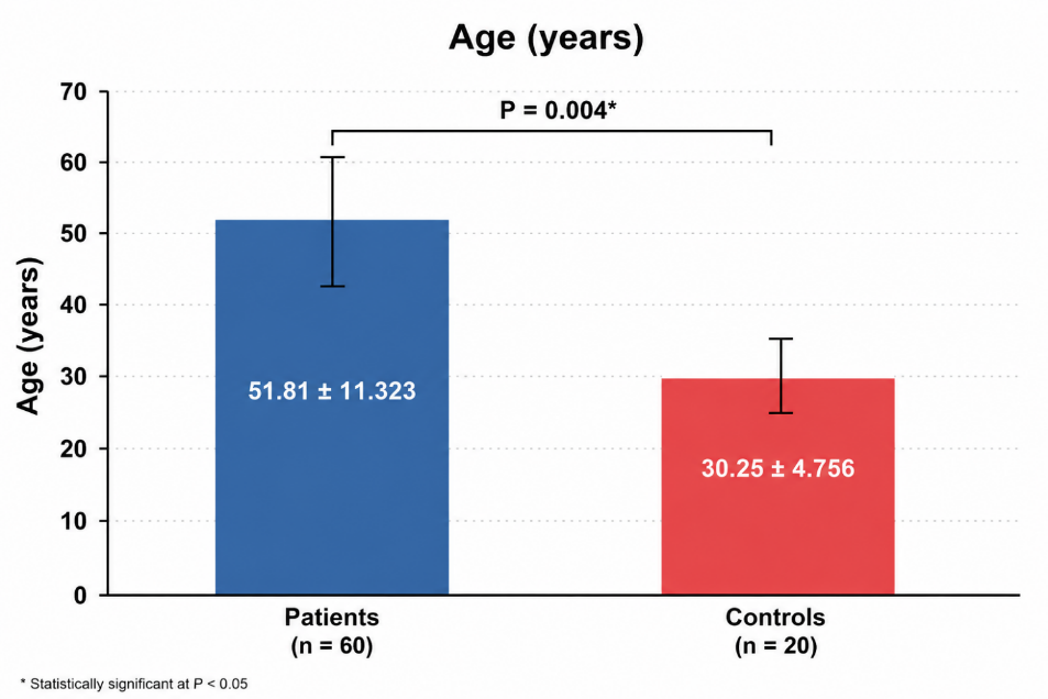
Figure 1: Compared the Mean of Age Between Breast Cancer Patients and Healthy Controls
Table 1: Summary of Significant Clinical Findings in Breast Cancer Patients
|
Parameter |
Patients (Mean±SD) |
Controls (Mean±SD) |
Change |
p-value |
|
CA15-3 (U/mL) |
23.44±10.35 |
16.61±7.02 |
↑ |
0.032 |
|
RBC (×10⁶/µL) |
4.44±0.65 |
4.80±0.62 |
↓ |
0.038 |
|
Hemoglobin (g/dL) |
12.52±1.33 |
13.89±1.44 |
↓ |
<0.001 |
|
ESR (mm/hr) |
34.57±19.56 |
15.12±9.30 |
↑ |
<0.001 |
|
ALP (U/L) |
94.70±28.52 |
67.80±18.59 |
↑ |
<0.001 |
|
Urea (mg/dL) |
28.30±11.03 |
23.09±7.06 |
↑ (Borderline) |
0.051 |
.
The current study also divided patients according to their age into three age groups: 25-45, 46-56 and 57-77; where 31% of patients were within 25-45 age group, 35% in the 46-56 group and 33.3% in 57-77 group, The majority of BC patients belonged to the 46–56 years age group (35.0%), followed by the 57–77 years (33.3%) and 25–45 years (31.7%) groups. This distribution suggests that BC occurred more frequently among middle-aged and older women in the present study, as presented in Figure 2.
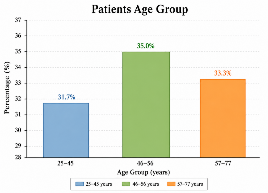
Figure 2: Distribution of Breast Cancer Patients According to Age Groups
The current investigation demonstrated that age of women with BC was significantly (p = 0.004) higher than that of the healthy individuals, where this finding is compatible with global epidemiological evidence indicated that the incidence of BC increasing with advancing age, exceptionally after fourth decade of life, since the cumulative genetic mutations, hormonal alterations and protracted exposure for environmental risk factors being collectively contributed to carcinogenesis [25]. Several reports indicated that nearly 80% of women with breast cancer at age >50, at that time, more than 40% were those with age >65 years [26-28]. Also, our findings align with several reports that show the incidence of breast cancer is peaking in women aged 45-65 years [29,30].
One of the most important factors contributing to BC in women over fifty is menopause, which represents a critical transformation in the health of women, as a result of the decline in ovarian function and hormonal fluctuations that contribute to different clinical issues, including metabolic alterations, weight gain and escalated insulin resistance, all of which might contribute to carcinogenesis.
Interestingly, later age at menopause extends the cumulative exposure to estrogen and progesterone, thus escalating the risk of breast tissue proliferation alongside malignant transformation. This interpretation is in line with the study of Hoinoiu et al. [31], which underscored the menopausal status as a key determinant of recurrence risk; they revealed that postmenopausal women often present with a distinct clinical pattern and comorbidities that complicate management outcomes. Similarly, a recent large case-control investigation revealed that postmenopausal women displayed notably higher BC risk as compared with premenopausal women, emphasizing the effect of hormonal exposure duration [32].
Regarding to distribution of patients according to age groups, the relatively balanced distribution of those points out the wide age spectrum influenced by BC, despite the predominance in middle-aged group (46-56) is consistent with global epidemiological data indicating that occurrence of breast cancer peaks throughout the premenopausal and early postmenopausal ages. Jacob et al. [32], demonstrated that women in this period experiences considerable hormonal fluctuation, especially dropped levels of estrogen and progesterone, which can engage with tumor development.
Moreover, the presence of a substantial proportion of younger patients (their ages ranging between 25-45 years) necessitates greater awareness of this disease risk, which is reported to be associated with more aggressive tumor status and poorer prognosis compared to those diagnosed later in life [30].
Assessment of CA15-3 Marker in Breast Cancer Patients and Healthy Individuals
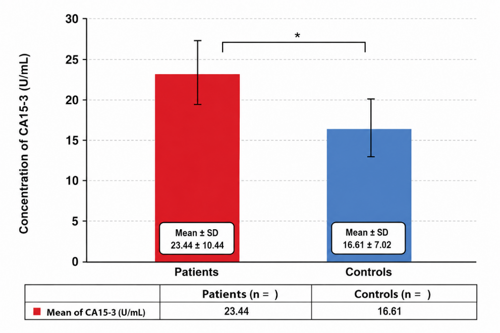
Patients with breast cancer had significantly higher serum CA15-3 concentrations than controls who were healthy (23.44±10.35 vs. 16.61±7.02 U/mL, p = 0.032, Z-test). The clinical relevance of CA15-3 as a helpful biomarker for disease identification and surveillance is supported by this notable spike, which shows elevated tumor-associated antigen gene expression in breast cancer-related patients.
The comparatively high standard deviations, however, point to inter-individual variation in CA15-3 levels among the participants under investigation.
Figure 3 displays serum concentrations of CA15-3 in studied population, where the present study revealed a significant (p = 0.032) increase of CA15-3 in women with BC versus healthy women. To measure some hematological parameters (including WBSs, RBCs, Hb, Platelets and ESR), the samples have been collected in EDTA and Hemolyzer Analyticon (Germany) automatic blood analysis device was employed to assess these parameters, this device takes the samples and automatically dilutes them, then the results are read.
.
Figure 3: Serum Concentrations of CA15-3 for Breast Cancer Patients and Healthy Controls
Results presented as mean±Standard Deviation (SD): 23.4443 ±10.3492 vs. 16.6050±7.0162, p = 0.032*, Z-test
The present study showed a significant increase of the CA15-3 marker in BC patients versus healthy women. This finding is aligned with previous reports that have emphasized this marker-3 as one of the most widely utilized tumor-associated antigens in patients with BC, especially to monitor disease progression and therapeutic responses rather than for early diagnosis [33,34]. This result is also in line with Assad et al. [35], who revealed that this transmembrane glycoprotein mucin is overexpressed in a number of epithelial malignancies, it possesses a considerable role in BC progression. Similarly, the Egyptian study conducted by Mohammed et al. [36], showed that patients with BC exhibited raised levels of CA15-3. Also, the findings of our study in line with studies conducted by Hamdi et al. [37] and Li et al. [38], which revealed that this tumor marker is abnormally elevated in patients with BC, suggesting a chance for disease progression or recurrence.
Additionally, our finding supported by the findings of Al-Saeedi et al. [39], who demonstrated a significant increase in CA15-3 in the serum of breast cancer patients without treatment and those receiving a single dose, compared to the control group. They suggested that CA15-3 as key requirement for diagnosing cases of breast cancer.
Assessment of Some Hematological Parameters in Breast Cancer Patients and Healthy Individuals
When compared to healthy controls, breast cancer patients showed significantly lower RBC counts and hemoglobin levels together with a noticeably higher ESR, which is indicative of anemic and systemic inflammation linked to cancer. WBC and platelet counts showed no discernible variations, suggesting that these hematological factors stayed largely constant in the sample under study.
Figure 4, displays the values of some hematological parameters (including WBSs, RBCs, Hb, Platelets and ESR) in in breast cancer patients and healthy controls. The results of statistical analysis showed only significant differences in RBCs, Hb and ESR; where RBCs and Hb were significantly decrease in breast cancer patients compared with control, while ESR values were significantly increase in patients.
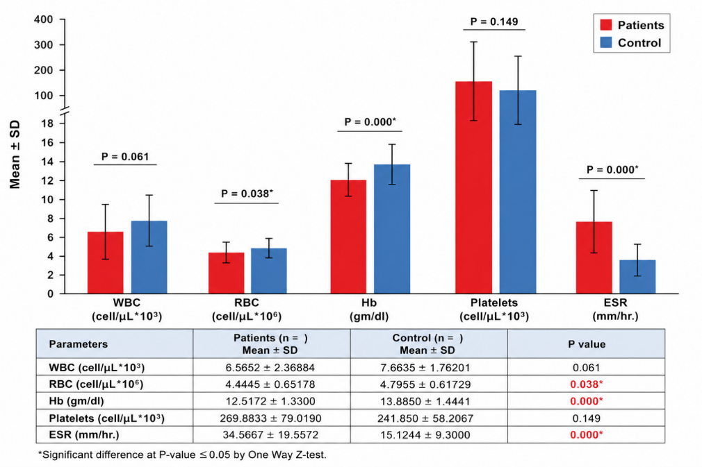
Figure 4: Assessment of Some Hematological Parameters in Breast Cancer Patients and Healthy Controls
The comparative analysis of hematological parameters demonstrates that significant changes were observed in RBCs, Hb and ESR values. In particular, RBCs and Hb levels were significantly decreased in breast cancer patients, while ESR values were significantly increased.
Hematological parameters are a reflection of cell-mediated immune responses against cancer and any change in these parameters affects the progression of cancer. Thus, the assessment of hematological parameters is dependable for both prognosis and diagnosis of various kinds of cancer, including breast cancer [40]. There is a demand for more research to comprehend the relationship between hematological parameters and breast cancer prognosis. Diminished Hb levels had been linked with poor prognosis and reduced treatment tolerance, underlining its clinical significance [41].
The present findings are in line with previous reports revealing that anemia is a common hematological abnormality in women with breast cancer, often attributed to chronic diseases, nutritional deficiency, or the myelosuppressive impacts of cancer-linked inflammations [11,42,43].
On the other hand, an Iranian study conducted by Rajizadeh et al. [44], showed non-significant differences in Hb and RBCs levels, whereas the values of MCV, MCH and MCHC were statistically significant.
In Turkey, Velidedeoglu et al. [45], found that white blood cells, neutrophils, lymphocytes and monocytes were significant, while hemoglobin, platelets and eosinophils were non-significant. Collectively, the hematological changes emphasized the interaction between cancer biology and the patient's systemic responses, suggesting that traditional observation of these hematological markers may offer valuable information in the clinical assessment and management of breast cancer patients.
Assessment of Liver Function Tests in Breast Cancer Patients and Healthy Individuals
Serum ALP levels were substantially higher in breast cancer patients than in healthy controls, although ALT and AST activities were not statistically different. These results imply that whereas routine hepatocellular enzymes stayed within similar ranges between the two groups, ALP may represent disease-related metabolic changes. In the term of liver function tests, the present study assesses the levels of three dependable liver function tests that included AST, ALT and ALP as presented in Figure 5.
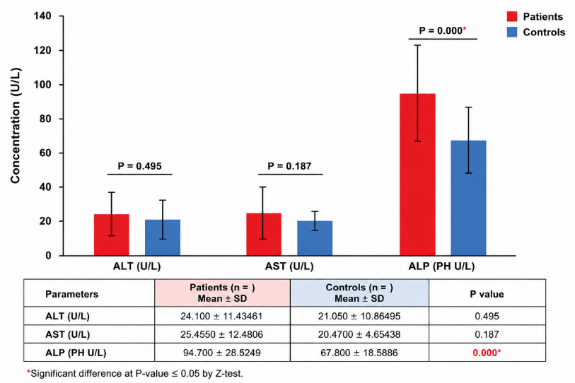
Figure 5: Assessment of Liver Function Tests in Breast Cancer Patients and Healthy Controls
The results of statistical analysis showed only a significant outcome with regard to ALP only, where its levels were significantly (p = 0.000) increase in women with breast cancer patients as compared with control women; while AST and ALT showed non-significant differences.
In the present study, liver function tests demonstrated a significant increase in ALP levels in breast cancer women compared to healthy individuals, while AST and ALT did not reveal significant differences. According to our results, the high ALP levels recorded in patients indicated that cancer had metastasized to the liver or bone. Alkaline phosphatase is a serum enzyme that reflects the integrated activity of a number of isoenzymes present in the liver, bone, kidney and epithelium of the intestine [46].
The roles of ALP in diagnosis, prognosis and surveillance of breast cancer should not be despised, especially in third-world populations that lack medical facilities or resource-poor settings [47]. Based on the biological activity of ALP enzyme, it has consistently been revealed to predict bone metastases and to some extent hepatic metastases [48,49]. High ALP levels may mirror increased osteoblastic activity as a result of skeletal metastases, which are common in developed breast cancer cases, or cholestatic alterations related to hepatic infiltration [50].
Our findings agree with Sing et al. who revealed a significant increase in ALP levels in different stages of cancer. Moreover, the present study is in line with Kh et al. [51], who reported high ALP levels in breast cancer patients as compared with healthy individuals; they also showed that ALP levels were raised significantly as the stage of cancer progressed. Additionally, our finding aligns with AL-Mashhadani et al. [52], who demonstrated that patients with bone metastases showed a significant increase in the levels of ALP. However, the findings of the current study partially agree with the recent study of Altimmime et al. [53], which revealed that the levels of ALP, ALT and AST were significantly increased in breast cancer women with metastasis as compared with patients without metastasis.
Assessment of Kidney Function Tests and Electrolytes in Breast Cancer Patients and Healthy Individuals
Serum urea levels were marginally higher in breast cancer patients (28.30±11.03 vs. 23.09±7.06 mg/dL) than in healthy controls, although this difference was not statistically significant (p = 0.051). In a similar vein, the two groups' serum creatinine levels were similar (p = 0.760). Serum electrolyte levels, such as Sodium (Na⁺), Potassium (K⁺) and Chloride (Cl⁻), showed no significant differences (p-values of 0.167, 0.271 and 0.871, respectively). These results show that the individuals with breast cancer under study had generally maintained electrolyte balance and renal function. Regarding the assessment of kidney function tests (urea and creatinine) and some electrolytes (Cl-, K+, Na+ mmol/L), the results of statistical analysis found that only urea is significant (p = 0.054) increase in patients, while creatinine and all studied electrolytes showed non-significant differences between patients and control as illustrated in Figure 6.
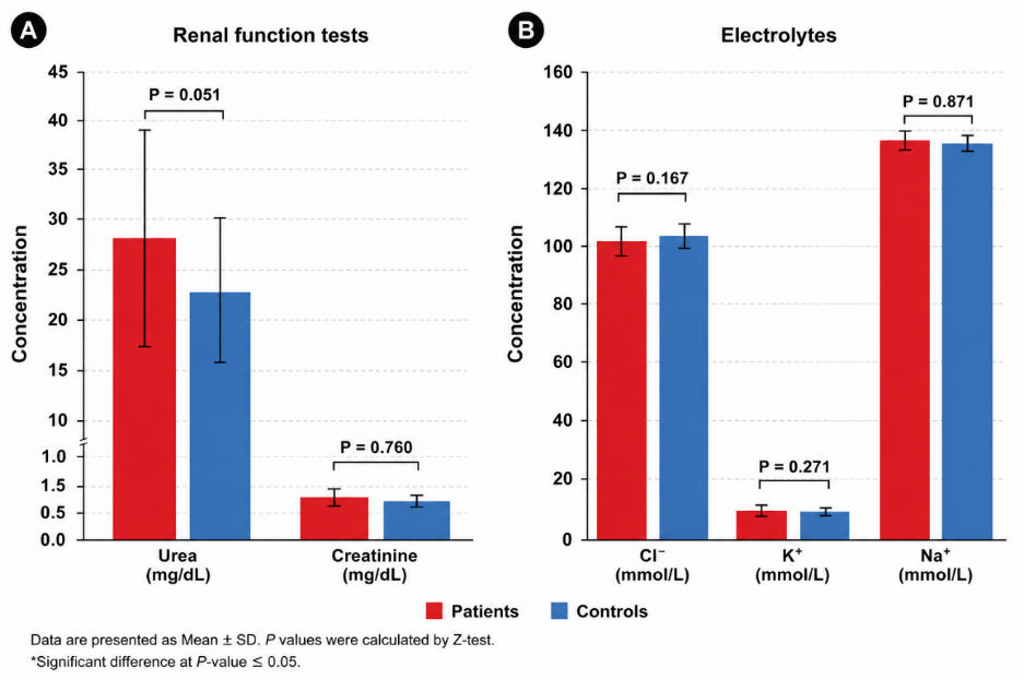
Figure 6: Comparison of Renal Function Tests and Serum Electrolyte Levels Between Breast Cancer Patients and Healthy Controls (A) Serum Urea and Creatinine Concentrations (mg/dL) in Patients and Controls. (B) Serum Electrolyte Concentrations, Including Chloride (Cl⁻), Potassium (K⁺) and Sodium (Na⁺) (mmol/L), in Both Groups. Data are Presented as Mean±Standard Deviation (SD)
The assessment of kidney function tests in our cohort demonstrated non-significant changes in sera levels of creatinine or electrolytes (Cl⁻, K⁺, Na⁺) between breast cancer women and healthy women, while urea levels displayed a borderline significant increase (p = 0.051) in patients. This pattern is consistent with a previous study that reported that kidney function is commonly preserved in breast cancer patients unless complicated by chemotherapy-linked nephrotoxicity or renal metastatic involvement [39].
This elevation of urea noticed in our cohort may reflect escalated protein catabolism and metabolic turnover correlated with malignancy, since cancer cells trigger a hypercatabolic state that raises the nitrogenous waste products [11]. Our findings are aligning with those of Chauhan et al. [54] and AL- Hussein [55], whose studies demonstrated a significant increase in urea levels in breast cancer patients compared to controls. Apart from toxicity of different therapies for these cancers, renal members are especially vulnerable for pre-existing comorbidities or other risk factors increasing the risk of renal impairment in patients prior to chemotherapy/radio therapy [56]. Chronic renal disease is common among the majority of elderly subjects in the general population, regardless of any cancer Stevens et al. [57].
The present study results were found to agree with Chauhan et al. [54], which showed that there is no significant difference in creatinine levels, with the average concentration is in the normal range for different individuals, including breast cancer patients and healthy subjects.
The results of the current study were consistent with the results of Al-Dulaimi, which stated that there were no significant differences in the serum creatinine level of breast cancer patients and healthy women. However, results of the current study were found to be different from the study held in Salahudin governorate in Iraq by Al-Hussein [55] and also differ with finding of the study by Devi et al. [58], indicated increased creatinine levels in women with breast cancer as compared to healthy women.
The Correlation Analysis
Only the count of WBC in breast cancer patients and the serum potassium levels in normal healthy controls showed a significant inverse link with CA15-3, no significant correlations were found with the other renal, hepatic, or electrolytic markers. These findings suggest that serum CA15-3 may function as a tumor-specific diagnostic in breast cancer and is comparatively independent of standard laboratory indicators. To determine the extent of CA15-3 correlation with all studied parameters in both patients and control groups, the current study performed a correlation analysis and showed that CA15-3 has a significant inverse correlation with WBCs in breast cancer patients and a significant inverse correlation with K+ only. No other significant correlations were found with the other studied parameters in either patients or healthy individuals as illustrated in Figure 7.
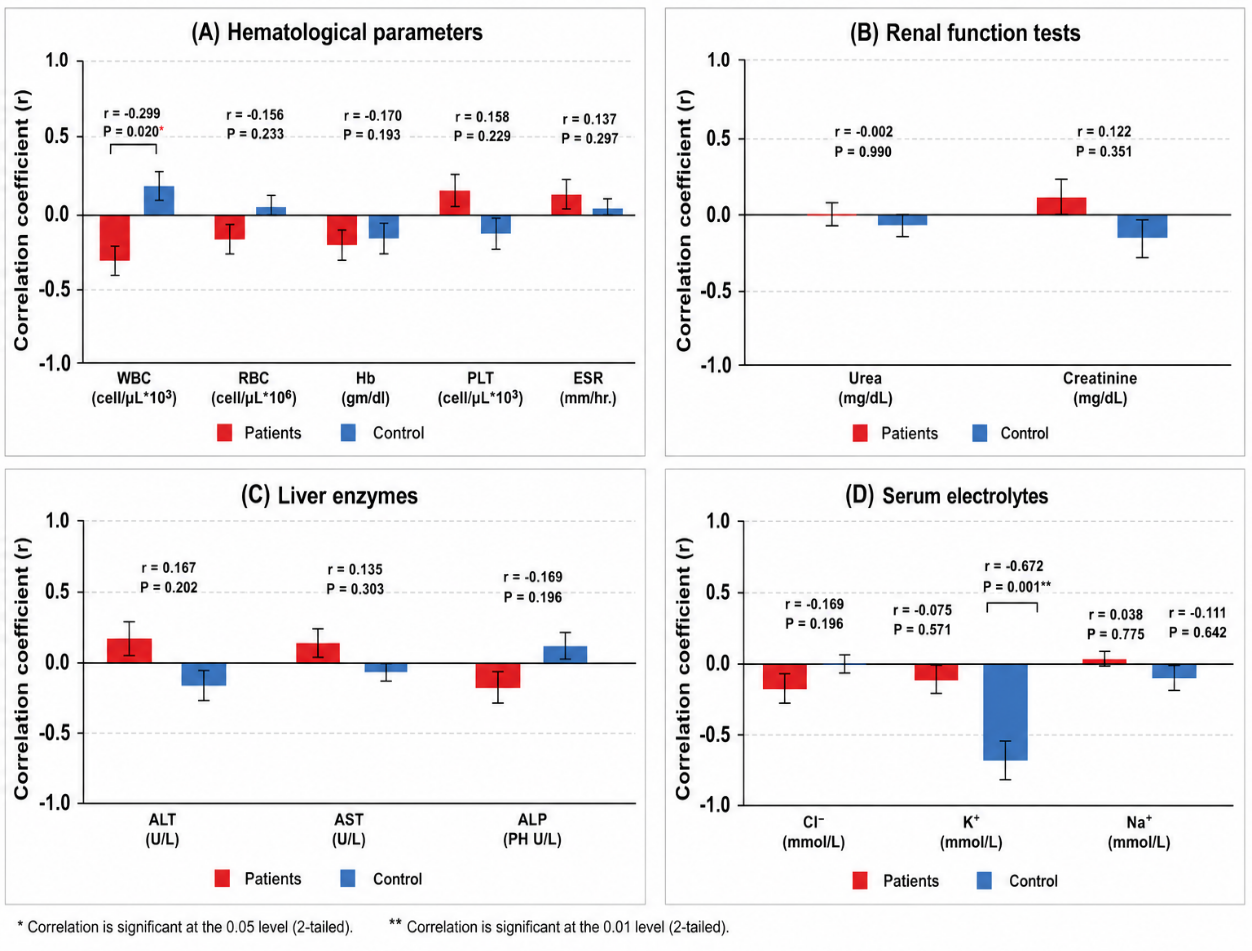
Figure 7: Correlation Between Serum CA15-3 Levels and Hematological, Renal, Hepatic and Electrolyte Parameters in Breast Cancer Patients and Healthy Controls. (A) Correlation of Serum CA15-3 with Hematological Parameters, Including White Blood Cell (WBC) Count, Red Blood Cell (RBC) Count, Hemoglobin (Hb), Platelet (PLT) Count and Erythrocyte Sedimentation Rate (ESR). (B) Correlation of Serum CA15-3 with Renal Function Markers (Urea and Creatinine). (C) Correlation of Serum CA15-3 with Liver Enzymes, Including Alanine Aminotransferase (ALT), Aspartate Aminotransferase (AST) and Alkaline Phosphatase (ALP). (D) Correlation of Serum CA15-3 with Serum Electrolytes (Cl⁻, K⁺ and Na⁺)
The correlation analysis demonstrates that CA15-3 is not only an indicator for the presence of cancer but it is also significantly associated with systemic immune responses and metabolic status of patients. The significant negative correlation between CA15-3 and WBC counts in patients aligns with the broader understanding of cancer-associated immune modulation; as tumor burden escalates (reflected by increasing CA15-3), the resulting chronic inflammations often lead to suppressed bone marrow or sequestration of leukocytes inside the tumor's microenvironment [59,60].
This phenomenon is termed immune exhaustion, the hallmark of advanced tumors where upraised tumor markers correspond with a decrease in effective circulating immune cells. Moreover, the significant negative correlation with K+ probably reflects metabolic stress and cellular turnover correlated with progressive pathology. According to Kumari et al. [61], about electrolyte imbalances in cancer, high-turnover states could disrupt Na+/K+-ATPase pump activity and shift the transcellular, leading to diminished serum K+ as a result of compromised cellular integrity. The non-correlative nature with liver and kidney indices and other electrolytes implies that CA15-3 is largely unaffected by global glomerular filtration rates and systemic osmotic balance, thereby reinforcing its nature predominantly as a cellular shed biomarker as opposed to a remote organ failure product [62].
CONCLUSIONS
The present study demonstrates that breast cancer is associated with distinct clinical and biochemical alterations. Patients exhibited significantly elevated serum CA15-3, ESR and ALP levels, together with reduced RBC count and hemoglobin, reflecting tumor-associated inflammation, anemia and increased tissue turnover. In contrast, liver transaminases (ALT and AST), creatinine and serum electrolyte levels remained largely unchanged, while urea showed only a borderline increase, indicating that renal function was generally preserved. Furthermore, CA15-3 displayed a significant inverse correlation with WBC count in breast cancer patients and with serum potassium levels in healthy controls, suggesting a potential association between tumor burden, immune response and metabolic regulation. Collectively, these findings support the value of combining CA15-3 with routine hematological and biochemical parameters to provide complementary information for the clinical assessment and monitoring of breast cancer, although further large-scale prospective studies are warranted to validate their prognostic and clinical utility.
REFERENCES
- World Health Organization (WHO). Breast Cancer: Prevention and Control. Geneva, 2024.
- International Agency for Research on Cancer (IARC). Global Cancer Observatory: Cancer Today. 2025.
- Freihat, O. et al. “Global burden and projections of breast cancer incidence and mortality to 2050: A Comprehensive Analysis of GLOBOCAN Data.” Frontiers in Public Health, vol. 13, 2025, article 1622954.
- Sung, H. et al. “Global cancer statistics 2020: Globocan estimates of incidence and mortality worldwide for 36 cancers in 185 countries.” CA: A Cancer Journal for Clinicians, vol. 71, no. 3, 2021, pp. 209–249.
- Wilkinson, L. and T. Gathani. “Understanding breast cancer as a global health concern.” The British Journal of Radiology, vol. 95, no. 1130, 2022, article 20211033.
- United Nations Development Programme (UNDP). Human Development Data. United Nations Development Programme, 2020.
- Gersten, O. and J.R. Wilmoth. “The cancer transition in Japan since 1951.” Demographic Research, vol. 7, 2002, pp. 271–306.
- Abdel, R.O. “The epidemiologic transition: A theory of the epidemiology of population change.” Bulletin of the World Health Organization, vol. 79, no. 2, 2021, pp. 161–170.
- Ali, L.O. “Study effect of breast cancer on some hematological and biochemical parameters in Babylon province, Iraq.” Journal of Pharmacy and Biological Sciences, vol. 9, no. 3, 2014, pp. 20–24.
- Madhu, Y. et al. “Hematological and biochemical serum markers in breast cancer: Diagnostic, therapeutic and prognostic significance.” Exploratory Research and Hypothesis in Medicine, vol. 10, no. 4, 2025.
- Madeddu, C. et al. “Pathogenesis and treatment options of cancer related anemia: Perspective for a targeted mechanism-based approach.” Frontiers in Physiology, vol. 9, 2018, article 1294.
- Ryu, J.M. et al. “prognostic impact of elevation of cancer antigen 15-3 (CA15-3) in patients with early breast cancer with normal serum CA15-3 level.” Journal of Breast Cancer, vol. 26, no. 2, 2023, pp. 126.
- Thompson, J.A. et al. “Carcinoembryonic antigen gene family: Molecular biology and clinical perspectives.” Journal of Clinical Laboratory Analysis, vol. 5, no. 5, 1991, pp. 344–366.
- Gendler, S.J. “MUC1, the renaissance molecule.” Journal of Mammary Gland Biology and Neoplasia, vol. 6, no. 3, 2001, pp. 339–353.
- Roy, L.D. et al. “MUC1 enhances invasiveness of pancreatic cancer cells by inducing epithelial to mesenchymal transition.” Oncogene, vol. 30, no. 12, 2011, pp. 1449–1459.
- Swapna, V.S. et al. “Study of liver function tests in breast carcinoma patients before and after chemotherapy.” International Journal of Biotechnology and Biochemistry, vol. 14, no. 3, 2018, pp. 177–184.
- Berardi, R. et al. “Electrolyte disorders in cancer patients: A systematic review.” Journal of Cancer Metastasis and Treatment, vol. 5, 2019.
- Leser, C. et al. “Liver function indicators in patients with breast cancer before and after detection of hepatic metastases: A retrospective study.” PLOS ONE, vol. 18, no. 3, 2023, article e0278454.
- Zhao, J. et al. “Global trends in incidence, death, burden and risk factors of early-onset cancer from 1990 to 2019.” BMJ Oncology, vol. 2, no. 1, 2023.
- Al Mashhadani, A.Z. et al. “Evaluation of G-ST, GOT and GPT activities in sera of leukemia patients before and after treatment.” Journal of Kerbala University, vol. 9, no. 3, 2011.
- Abdullah, S.O. et al. “Study of liver function test and vitronectin as biomarker for liver cell in Iraqi chronic myeloid leukemia (CML) patients with nucleophosmin 1 (NPM) mutation.” Research Journal of Biotechnology, vol. 19, no. 11, 2024, pp. 165–171.
- Al-Musawi, H.S. et al. “Age and gender impact on glycaemic control, renal function and oxidative stress parameters in iraqi patients’ type 2 diabetes mellitus.” Biochemical & Cellular Archives, vol. 21, no. 1, 2021a.
- Al-Musawi, H.S. et al. “Age and gender impact on glycaemic control, renal function and oxidative stress parameters in Iraqi patients type 2 diabetes mellitus.” Biochemical & Cellular Archives, vol. 21, no. 1, 2021b.
- Sakhi, H.S. et al. “The study of some biochemical markers associated with prognosis of breast cancer in Iraqi women.” Journal of Kerbala University, vol. 21, no. 1, 2024.
- Łukasiewicz, S. et al. “Breast cancer epidemiology, risk factors, classification, prognostic markers and current treatment strategies: An updated review.” Cancers, vol. 13, no. 17, 2021, article 4287.
- Benz, C.C. “Impact of aging on the biology of breast cancer.” Critical Reviews in Oncology/Hematology, vol. 66, no. 1, 2008, pp. 65–74.
- Siegel, R. et al. “Cancer statistics, 2014.” CA: A Cancer Journal for Clinicians, vol. 64, no. 1, 2014.
- McGuire, A. et al. “Effects of age on the detection and management of breast cancer.” Cancers, vol. 7, no. 2, 2015, pp. 908–929.
- Bray, F. et al. “Global cancer statistics 2018: GLOBOCAN estimates of incidence and mortality worldwide for 36 cancers in 185 countries.” CA: A Cancer Journal for Clinicians, vol. 68, no. 6, 2018, pp. 394–424.
- DeSantis, C.E. et al. “Breast cancer statistics, 2019.” CA: A Cancer Journal for Clinicians, vol. 69, no. 6, 2019, pp. 438–451.
- Hoinoiu, T. et al. “Risk factors for breast cancer recurrence in postmenopausal women: A bibliometric study.” Frontiers in Oncology, vol. 15, 2025, article 1522713.
- Jacob, J.S. et al. “Unraveling breast cancer risk with a focus on menopausal status: A large case–control study.” Cancer Causes & Control, vol. 37, 2026, pp. 15.
- Sturgeon, C.M. et al. “National academy of clinical biochemistry laboratory medicine practice guidelines for use of tumor markers in testicular, prostate, colorectal, breast and ovarian cancers.” Clinical Chemistry, vol. 54, no. 8, 2008, pp. e1–e10.
- Khadhum, H.S. et al. “Assessment of CA 15-3 and P53 biomarkers in diagnosis of breast cancer.” Assessment, vol. 140, no. 1, 2022.
- Assad, D.X. et al. “Correlation between salivary and serum CA15-3 concentrations in patients with breast cancer.” Molecular and Clinical Oncology, vol. 13, no. 2, 2020, pp. 155–161.
- Mohammed, F.Z. et al. “Assessment of CA15.3 and CEA as potential markers for breast carcinoma prognosis in egyptian females.” Alfarama Journal of Basic & Applied Sciences (AJBAS), vol. 2, no. 1, 2021, pp. 44–50.
- Hamdi, E.T. et al. “The relationship between vitamin d with breast cancer.” Medical Science, vol. 24, no. 104, 2020, pp. 2600–2603.
- Li, J. et al. “Tumor markers CA15-3, CA125, CEA and breast cancer survival by molecular subtype: A cohort study.” Breast Cancer, vol. 27, no. 4, 2020, pp. 621–630.
- Al-Saeedi, T.K.J. et al. “Evaluation CA15-3, blood urea and creatinine in breast cancer women.” Iraqi National Journal of Chemistry, vol. 23, no. 1, 2023, pp. 1–11.
- Divsalar, B. et al. “Hematological parameters changes in patients with breast cancer.” Clinical Laboratory, no. 8, 2021.
- Knight, K. et al. “Prevalence and outcomes of anemia in cancer: A systematic review of the literature.” The American Journal of Medicine, vol. 116, no. 7, 2004, pp. 11–26.
- Udosen, J.E. et al. “Some hematological parameters of breast cancer patients accessing therapy at University of Calabar teaching hospital, Calabar Nigeria.” Sokoto Journal of Medical Laboratory Science, vol. 7, no. 1, 2022.
- Abbas, A.B. et al. “Comparison of hematological parameters and the associated factors among women with and without breast cancer: A case-control study.” Breast Cancer (Dove Medical Press), vol. 16, 2024, pp. 877–885.
- Rajizadeh, A. et al. “Comparison of hematological parameters, iron levels and oxidative stress in women with and without breast cancer: A case-control study.” Medical Journal of the Islamic Republic of Iran, vol. 31, 2017, article 114.
- Velidedeoglu, M. et al. “Preoperative fibrinogen and hematological indexes in the differential diagnosis of idiopathic granulomatous mastitis and breast cancer.” Medicina, vol. 57, no. 7, 2021, article 698.
- Keshaviah, A. et al. “CA15-3 and alkaline phosphatase as predictors for breast cancer recurrence: A combined analysis of seven international breast cancer study group trials.” Annals of Oncology, vol. 18, no. 4, 2007, pp. 701–708.
- Singh, S. et al. “Recent update on serum alkaline and acid phosphatases in pre- and postoperative breast cancer patients.” Sudan Journal of Medical Sciences, vol. 17, no. 1, 2022, pp. 70–78.
- Reichling, J.J. and M.M. Kaplan. “Clinical use of serum enzymes in liver disease.” Digestive Diseases and Sciences, vol. 33, no. 12, 1988, pp. 1601–1614.
- Friedman, L.S. et al. “Liver function tests and the objective evaluation of the patient with liver disease.” Hepatology: A Textbook of Liver Disease, vol. 1, 1996, pp. 791–833.
- Nicolini, A. et al. “Prognostic and predictive biomarkers in breast cancer: Past, present and future.” Seminars in Cancer Biology, vol. 52, 2018, pp. 56–73.
- Kh, C. et al. “Significance of serum total alkaline phosphatase levels in breast cancer.” International Journal of Clinical and Biomedical Research, vol. 2, no. 1, 2016, pp. 13–15.
- AL-Mashhadani, Z.I. et al. “Estimation of ALP, GPT and GOT activities in iraqi patients female with breast cancer.” Ibn AL-Haitham Journal for Pure and Applied Science, vol. 25, no. 1, 2017.
- Altimmime, B.A. et al. “CXCL12, CA15-3 and liver function tests as predictors of liver metastasis in breast cancer.” Al-Nahrain Journal of Science, vol. 28, no. 4, 2025, pp. 55–66.
- Chauhan, P. et al. “Evaluation of serum biochemical profile of breast cancer patients.” International Journal of Medical Research & Health Sciences, vol. 5, no. 7, 2016, pp. 1–7.
- AL-Hussein, S.A.H. “Evaluation of the serum levels of alanine aminopeptidase and aggrecan with some biochemical parameters in women with breast cancer.” Indian Journal of Forensic Medicine & Toxicology, vol. 14, no. 3, 2019, pp. 2535–2540.
- Aapro, M. and V. Launay-Vacher. “Importance of monitoring renal function in patients with cancer.” Cancer Treatment Reviews, vol. 38, no. 3, 2012, pp. 235–240.
- Stevens, L.A. et al. “Prevalence of CKD and comorbid illness in elderly patients in the United States: Results from the kidney early evaluation program (KEEP).” American Journal of Kidney Diseases, vol. 55, no. 3, 2010, pp. S23–S33.
- Devi, L.I. et al. “Serum biochemical profile of breast cancer patients.” European Journal of Pharmaceutical and Medical Research, vol. 2, no. 6, 2015, pp. 210–214.
- Ri, G. et al. “Serum levels of CA15-3, KL-6 and BCA225 Are positively correlated with each other in the general population.” Anticancer Research, vol. 29, no. 10, 2009, pp. 4239–4242.
- Duffy, M.J. “CA 15-3: Uses and limitations as a biomarker for breast cancer.” Clinical Chemistry, vol. 54, no. 12, 2008, pp. e11–e79.
- Siddiqui, A.A. et al. “Correlation of duration of chemotherapy with electrolytes in cancer patients: A prospective study assessing the relationship with various electrolytes.” Age (Years), 2018, pp. 12–81.
- Hing, J.X. et al. “Clinical utility of tumor marker velocity of cancer antigen 15–3 (CA 15–3) and carcinoembryonic antigen (CEA) in breast cancer surveillance.” The Breast, vol. 52, 2020, pp. 95–101.